

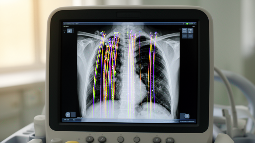
A prospective clinical investigation conducted at Skåne University Hospital in Sweden demonstrates that an automated artificial intelligence tool integrated into a commercial point-of-care ultrasound system substantially overcounts pulmonary B-lines compared to visual reviews performed by expert physicians. Lung ultrasound has become an essential bedside examination for evaluating pulmonary edema and interstitial lung syndrome through the identification of B-lines, which are vertical, hyperechoic, laser-like artifacts that originate from the pleural line and extend across the entire imaging sector without fading. In this single-center evaluation of fifty-five hospitalized patients presenting with acute respiratory symptoms, primarily acutely decompensated heart failure, investigators found poor statistical agreement between the automated algorithm and experienced human evaluators, driven largely by the platform’s propensity to overestimate artifact counts across multiple anatomical thoracic zones.
Assessing the accumulation of extravascular lung water using lung ultrasound offers higher sensitivity than conventional chest radiography and carries key prognostic importance for tailoring diuretic therapy and preventing hospital readmission in decompensated heart failure. Because visual identification and scoring of B-lines typically demand specialized training and clinical experience, automated software solutions have been deployed by ultrasound manufacturers to standardize evaluations and assist non-expert operators. The software tested in this study was the automated B-line application embedded within the Venue GO point-of-care ultrasound platform manufactured by General Electric Healthcare. The commercial tool is designed to highlight and quantify B-lines in real time across captured video loops, ultimately logging the frame with the maximum artifact count to assign a score for each scanned lung zone.
The study protocol gathered recordings from adult inpatients admitted to the Internal Medicine and Cardiology departments who were able to sit upright and give informed consent. Patients with known pre-existing lung diseases or pneumonia were excluded from enrollment, although individuals with asthma or pulmonary embolism remained eligible. Examinations were executed at the bedside by an internal medicine resident undergoing point-of-care ultrasound training using a curved low-frequency transducer set to a standard imaging depth of sixteen centimeters. A standardized twelve-zone thoracic scanning protocol was implemented, consisting of four anterolateral zones recorded on each side of the chest with the patient inclined supine at forty-five degrees, followed by two dorsal zones recorded on each side of the back while the patient sat upright on the edge of the examination bed.
Each recorded sector was captured for four
Each recorded sector was captured for four to five seconds in the sagittal plane between adjacent ribs. To ensure consistent baseline image acquisition across all fifty-five subjects, a single operator acquired every video loop. The automated tool assigned each scanned zone an artifact category of zero, one, two, three, four, or greater than five B-lines, recording a total of 672 thoracic zone clips. Two independent physician experts, consisting of a cardiologist experienced in cardiac and lung ultrasound and an internal medicine consultant who directs a regional point-of-care ultrasound training academy, independently evaluated and classified B-lines into categorical brackets of zero, one to two, three to four, and five or more while remaining fully blinded to the software’s automated classifications.
Of the 672 recorded thoracic clips, 574 clips representing eighty-five percent of all acquired data were determined eligible for statistical analysis. Ninety-eight clips were excluded from the correlation assessment: eleven were deemed of insufficient image quality, twenty-six failed to demonstrate an identifiable pleural line, and fifty-one clips contained pleural effusions. Notably, twenty-two of the twenty-six clips lacking a clear pleural reflection occurred in the second left anterior zone, where cardiac motion directly obscured the pulmonary field. Pleural effusions were predominantly identified in the dependent posterior inferior zones, accounting for sixty-three percent of effusion findings, with twenty-nine percent appearing in the lateral chest regions.
Comparison of quantitative results revealed pronounced numerical discrepancies between automated assessments and human experts. Across all analyzed zones, the median B-line count tallied by the artificial intelligence platform was 23.5 per patient, compared to a median of 2.8 per patient determined by the visual reviewers, representing a statistically significant difference. The distribution of artifact categories demonstrated a strong skew in algorithmic scoring: the automated platform classified thirty-eight percent of all assessed zones into the highest category of more than five B-lines, whereas expert physicians assigned only four percent of the zones to that severe category. The automated software showed a bimodal distribution, placing forty-eight percent of readings at zero and thirty-eight percent at greater than five, with only thirteen percent falling between one and four B-lines.
Statistical agreement measured via two-way mixed-effects intraclass correlation
Statistical agreement measured via two-way mixed-effects intraclass correlation coefficient models indicated poor concordance between the automated tool and human experts. The intraclass correlation coefficient between the software and expert reviewers reached only 0.28 for the total patient B-line sum and 0.37 when assessed using a zone-by-zone comparison. In sharp contrast, interrater reliability between the two independent human expert physicians was excellent, yielding intraclass correlation coefficients of 0.92 for the total B-line sum and 0.91 for zone-by-zone scoring. Bland-Altman analysis similarly demonstrated a systematic positive bias of 1.86 with a standard deviation of 2.15 and wide ninety-five percent limits of agreement spanning from negative 2.35 to positive 6.07, illustrating frequent instances where the software scored five artifacts in sectors where clinicians observed zero.
Detailed qualitative inspection of all video clips where the software flagged one or more B-lines explained several technical failure modes contributing to algorithmic overcounting. Among the 574 analyzed zones, fourteen percent of algorithmic errors stemmed from the software tracking and evaluating acoustic data in the adjacent rib spaces rather than restricting analysis strictly to the central intercostal target, even though examiners maintained proper acoustic probe alignment. In one percent of evaluated zones, the software mistook subpleural consolidations displaying irregular acoustic shred lines, also designated C-lines, for true vertical B-lines. In another one percent of zones, normal acoustic rib shadowing positioned between intercostal spaces was incorrectly quantified as pathologic vertical artifacts.
Correlation between software and experts varied markedly by anatomical region, with the poorest correlation occurring in the anterior zone two and posterior superior zone five, which were the thoracic sectors where algorithmic overcounting was most pronounced. Better regional correlation was observed in the lateral and posterior inferior lung sectors. Clinical correlation checks confirmed that human expert counts reflected patient volume status, with reviewers recording significantly higher total B-line sums in hypervolemic patients compared to euvolemic individuals, averaging 4.6 versus 0.75 artifacts, respectively.
The study authors highlighted several methodological considerations
The study authors highlighted several methodological considerations and constraints, including recruitment from a single medical center, reliance on a single image operator, and the technical limitation of the embedded software capping granular counting at five artifacts per sector. While earlier small-scale publications involving as few as four patients had reported good software agreement with experts, the broader real-world inpatient sample evaluated in this trial mirrored the lower correlation figures documented in recent independent literature. The researchers concluded that automated B-line quantification embedded within current commercial point-of-care hardware requires further algorithmic refinement before it can be safely relied upon for autonomous clinical decision-making.
To align the differing categorization schemes during statistical analysis, researchers adjusted the automated software outputs, assigning values of one or two to 1.5 and values of three or four to 3.5 to mirror the expert score ranges. Beyond rib-space tracking errors, the investigators highlighted clinical diagnostic complexities rooted in ultrasound physics. Specifically, true cardiogenic B-lines generate smooth pleural reflections appearing in a diffuse pattern, whereas comet-tail artifacts arising from other pulmonary disorders feature irregular, fragmented pleural reflections with narrow heads and wide tails. The automated platform may struggle to differentiate between these distinct acoustic presentations, leading to inflated artifact tallies across heterogeneous patient groups.
The investigation also contextualizes earlier literature and promotional benchmarks associated with the commercial system. An initial evaluation by Short and colleagues cited in platform marketing materials documented an intraclass correlation coefficient of 0.79 using the same software, but that study evaluated only four patients. Later assessments revealed considerably lower concordance, such as an evaluation by Russell and colleagues that reported an intraclass correlation of 0.56 between automated scoring and expert review. Similarly, an investigation by Damodaran and colleagues examining patients with viral respiratory infections recorded intraclass correlation coefficients of 0.52 to 0.53, reflecting borderline moderate agreement that aligns much more closely with the modest correlation observed in the Swedish cohort.
Scanning protocols and architectural approaches also bear
Scanning protocols and architectural approaches also bear on clinical integration. The Swedish study implemented an upright twelve-zone method including dorsal regions, whereas recent heart failure research suggests a simplified two-zone anterior-superior thoracic protocol can yield comparable diagnostic utility to an eight-zone exam. Furthermore, current commercial point-of-care tools rely primarily on prespecified image-processing rules that struggle when encountering rib margins or tissue consolidations. The study authors noted that future improvements likely depend on deep-learning neural networks trained on extensive imaging datasets coupled with expert human visual validation to achieve the precision required for reliable point-of-care decision support.
Subject of Research: Evaluation of artificial intelligence software for quantifying pulmonary B-lines in bedside lung ultrasound
Article Title: Performance of a point-of-care ultrasound platform for artificial intelligence-enabled assessment of pulmonary B-lines
Article References: Labaf, A., Åhman-Persson, L., Husu, L
Article References: Labaf, A., Åhman-Persson, L., Husu, L. S., Smith, J. G., Ingvarsson, A., & Evaldsson, A. W. (2025). Performance of a point-of-care ultrasound platform for artificial intelligence-enabled assessment of pulmonary B-lines. Cardiovascular Ultrasound, 23(1), Article 3. https://doi.org/10.1186/s12947-025-00338-2
DOI:10.1186/s12947-025-00338-2
Keywords: point-of-care ultrasound, lung ultrasound, artificial intelligence, pulmonary B-lines, heart failure, pulmonary congestion, extravascular lung water, intraclass correlation coefficient, Bland-Altman analysis, interrater reliability, clinical evaluation, thoracic imaging, interstitial lung syndrome, General Electric Healthcare, diagnostic accuracy, bedside imaging
News. Point-of-Care Ultrasound Artificial Intelligence Overcounts Pulmonary B-Lines Compared to Clinical Experts. Scienmag
